Article

Determinants of Functional Capacity and Quality of Life After Implantation of a Durable Left Ventricular Assist Device

Register or Login to View PDF Permissions
Permissions× For commercial reprint enquiries please contact Springer Healthcare: ReprintsWarehouse@springernature.com.

For permissions and non-commercial reprint enquiries, please visit Copyright.com to start a request.

For author reprints, please email rob.barclay@radcliffe-group.com.
Average (ratings)
No ratings
Your rating

Abstract

Continuous-flow left ventricular assist devices (LVAD) are increasingly used as destination therapy in patients with end-stage heart failure and, with recent improvements in pump design, adverse event rates are decreasing. Implanted patients experience improved survival, quality of life (QoL) and functional capacity (FC). However, improvement in FC and QoL after implantation is not unequivocal, and this has implications for patient selection and preimplantation discussions with patients and relatives. This article identifies preimplantation predictors of lack of improvement in FC and QoL after continuous-flow LVAD implantation and discusses potential mechanisms, allowing for the identification of potential factors that can be modified. In particular, the pathophysiology behind insufficient improvement in peak oxygen uptake is discussed. Data are included from 40 studies, resulting in analysis of >700 exercise tests. Mean peak oxygen uptake was 13.4 ml/ kg/min (equivalent to 48% of predicted value; 259 days after implantation, range 31–1,017 days) and mean 6-minute walk test distance was 370 m (182 days after implantation, range 43–543 days). Finally, the interplay between improvement in FC and QoL is discussed.

Disclosure:FG is a consultant for Abbott and has received speakers’ fees from Abbott and Carmat. KKM has no conflicts of interest to declare.

Received:

Accepted:

Correspondence Details:Finn Gustafsson, Department of Cardiology, Section 2142, Rigshospitalet, Copenhagen University Hospital, Blegdamsvej 9, DK-2100 Copenhagen, Denmark. E: Finn.Gustafsson@regionh.dk

Copyright Statement:

The copyright in this work belongs to Radcliffe Medical Media. Only articles clearly marked with the CC BY-NC logo are published with the Creative Commons by Attribution Licence. The CC BY-NC option was not available for Radcliffe journals before 1 January 2019. Articles marked ‘Open Access’ but not marked ‘CC BY-NC’ are made freely accessible at the time of publication but are subject to standard copyright law regarding reproduction and distribution. Permission is required for reuse of this content.

According to the third report from the International Society for Heart and Lung Transplantation (ISHLT) Mechanically Assisted Circulatory Support Registry, more than 15,500 left ventricular assist devices (LVADs) were implanted worldwide between 2013 and 2017.1 Originally, LVADs were exclusively implanted as part of a bridge to transplantation (BTT) strategy, but in recent years a large proportion of patients undergoing LVAD implantation received the device as final treatment for advanced heart failure (HF), so-called destination therapy.

Survival rates after LVAD implantation have increased greatly over the past decade, as improvements in LVAD technology and management have resulted in a lower risk of adverse events.1–7 Hence, in recent studies, 2-year survival rates range from 80% to 90%.5,8 Patients treated with LVADs experience significant improvements in HF symptoms, quality of life (QoL) and functional capacity (FC), although the latter remains reduced in this patient group, especially when measured as peak oxygen uptake (pVO2).3,6–11

Given the increased use of LVAD as destination therapy and the long wait times for transplantation in those implanted with an LVAD as BTT, optimisation of FC and QoL is critically important.1 In order to identify potential strategies to improve FC and QoL after LVAD implantation, a detailed understanding of the mechanisms behind residual impairment of these parameters is essential.

This review summarises the available evidence describing improvement in FC and QoL after LVAD implantation with the specific aim of identifying reproducible predictors of improvement in QoL and FC with the intervention.

Methods

Search Strategy

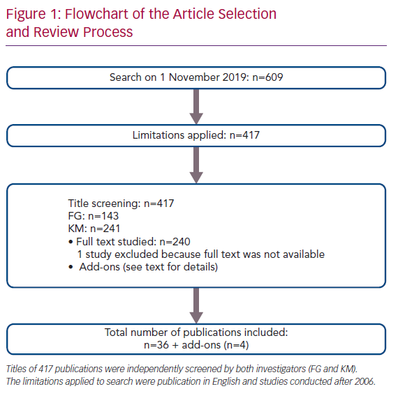
On 1 November 2019, a PubMed search was conducted using the search terms ‘quality of life’, ‘EuroQoL-5 Dimensions-5 levels’, ‘Minnesota Living With Heart Failure’, ‘Kansas City Cardiomyopathy Questionnaire’, ‘exercise’, ‘six minute walk test’, ‘six-minute walk test’, ‘peak oxygen uptake’, ‘exercise capacity’, ‘peak oxygen consumption’, ‘exercise training’, ‘cardiac rehabilitation’, ‘ventricular assist device’ and ‘continuous-flow left ventricular assist device’ (Supplementary Material Table 1). The search resulted in 609 items (Figure 1). After excluding studies published prior to 2006 and those written in languages other than English, the titles of 417 publications were screened by both investigators independent of each other (i.e. blinded). This process resulted in the identification of 143 publications by FG and 241 publications by KM. Mismatch and disagreement in 98 cases led to full-text review. The full text of one article could not be acquired, which resulted in its exclusion. Finally, 36 papers and four ‘add-on’ studies (e.g. extra studies found by reference review or other sources) were included in this narrative review.9,12–50

Flowchart of the Article Selection and Review Process

Article image

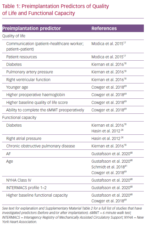
Preimplantation Predictors of Quality of Life and Functional Capacity

Article image

Calculations

Mean values weighted for population size for pVO2 and the 6-minute walk test (6MWT) were calculated as follows:

Weighted mean pVO2 = Σ(pVO2 × npVO2)/ΣNpVO2

Weighted mean 6MWT = Σ(6MWD × n6MWT)/ΣN6MWT

Where n refers to the number of patients in each study, while N is the total number of all studied patients. 6MWD is the distance covered in the 6MWT.

Results and Discussion

Functional Capacity After LVAD Implantation

The most widely accepted measures of functional capacity in HF are symptoms, measured by New York Heart Association (NYHA) class, 6MWD and pVO2. Each of these measures has its own advantages and disadvantages, such as variable reproducibility or technical demands, but all are useful and required to characterise different aspects of the exercise limitation of HF populations. Patients referred for LVAD implantation are almost invariably severely symptomatic (e.g. NYHA IIIb–IV with 6MWD <300–400 m and low pVO2). The latter is typically below 12 ml/kg/min, which is also the limit used as part of the indication for destination therapy LVAD in the US Medicare system.

In all studies of patients undergoing LVAD implantation, NYHA class improves in most patients from Class III/IV before implantation (100%) to Class I or II.9,49 Studies are remarkably consistent in finding that approximately 80% of patients are in NYHA Class I–II after implantation, and the improvement in symptoms has been documented to be sustained over time.9,20,49 However, some patients (<5%) remain severely symptomatic (NYHA Class IV) even 12 months after LVAD implantation.9,49

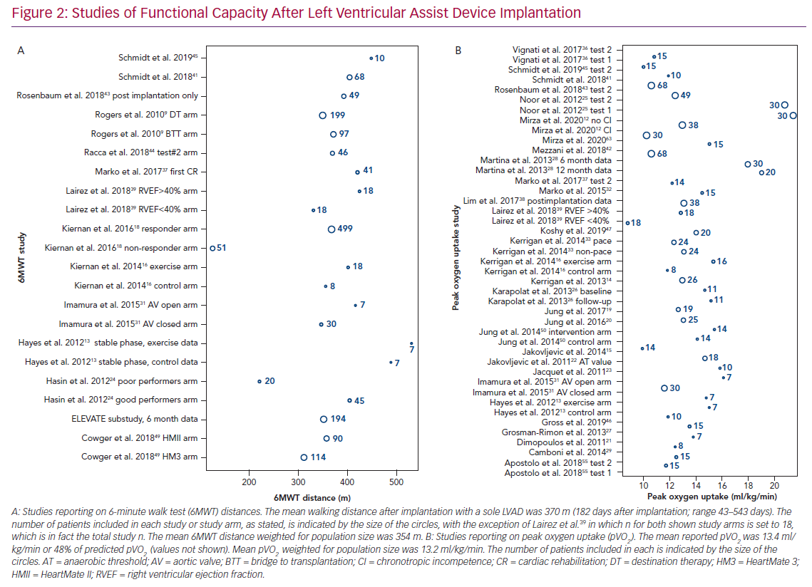
The 6MWD is widely used in LVAD recipients.9,13,16,24,31,48,49 FC measured using the 6MWT prior to implantation is low (mean 6WMD 221 m [range 39–356 m]; mean 6MWD weighted for population size 215 m), and significant improvements are observed soon after implantation (mean 6WMD 373 m [range 126–531 m]; mean 6MWD weighted for population size 357 m; Figure 2A).9,18,24,43,49 The improvement from baseline to the 6-month follow-up is, on average, +144 m (range 41–319 m; mean improvement weighted for population size 113 m), which is equivalent to an approximate 40% improvement.9,18,24,43,48,49

Improvement in the 6MWD can be difficult to interpret across different studies because some studies include mostly ambulatory patients and some include patients who would not be able to complete any exercise testing prior to implantation due to critical clinical condition (e.g. Interagency Registry of Mechanically Assisted Circulatory Support [INTERMACS] profile 1–2). In the large LVAD trials, increments in 6MWD were in the range 98–250 m from baseline to a maximum 2 years after implantation5,9,51–60 When investigating FC expressed as pVO2, the reported preimplantation values are low (mean 11 ml/kg/min [range 10.1–11.8 ml/kg/min]; mean pVO2 weighted for population size 11 ml/kg/min), although improvement is observed because studies reporting pre- and postimplantation pVO2 values show an improvement of approximately 20% after implantation.27,38,43

Postimplantation mean pVO2 values (Figure 2B) vary from 8.8 to 21.4 ml/kg/min (mean pVO2 weighted for population size 13.2 ml/kg/min), showing that pVO2 generally remains reduced after implantation at, on average, 48% of the expected value for age and sex.12–16,21–23,25–29,31–37,39–42,44–47,61–63 A considerable proportion of the variance of postimplantation pVO2 between published studies can be attributed to differences in the mean age of included patients.11

Studies of Functional Capacity After Left Ventricular

Article image

Although the improvement from pre- to postimplantation FC, measured as both 6MWT and pVO2, may appear modest, these changes are much larger than the effect of other device therapies in HF, such as cardiac resynchronisation therapy or the use of vasodilators.64,65 Furthermore, it should be re-emphasised that the sickest patients were excluded from studies presenting changes in FC from before to after implantation because these patients were not able to complete preimplantation measurements (e.g. because of the need for ventilator treatment or temporary mechanical circulatory support). Hence, the improvement in 6MWD and pVO2 from before to after implantation is often underestimated in the literature.

Preimplantation Predictors of Postimplantation Functional Capacity

Several studies have elucidated preimplantation determinants of postimplantation QoL and/or FC (Table 1 and Supplementary Material Table 2).17,18,24,27,38,43,48,49

Advanced age is generally a predictor of inferior outcomes in cardiovascular medicine; this also holds true for patients implanted with an LVAD. In a Multicenter Study of MAGLEV Technology in Patients Undergoing Mechanical Circulatory Support Therapy with HeartMate 3 (MOMENTUM 3) substudy including 265 patients from the US, younger age was one of the strongest predictors of the ‘living well on a left ventricular assist system’ endpoint (6MWD >300 m and Kansas City Cardiomyopathy [KCCQ] score >50).49 Confirming these findings, Martina et al. showed that HeartMate II (HMII) recipients <50 years of age performed significantly better than LVAD recipients >50 years of age.28 Several other studies have confirmed age as an independent preoperative determinant of postimplantation FC.41,46,48

Other strong preoperative predictors of FC are a lower INTERMACS profile, NYHA class, chronic obstructive pulmonary disease (COPD), diabetes and lower estimated glomerular filtration rate (eGFR).15,24,24 Higher haemoglobin, eGFR and INTERMACS profile and better NYHA class at the time of implantation all reflect a general better health status, which is linked to better FC after implantation. In general, these findings are suggestive of benefits, at least in terms of FC improvement, of early implantation of LVADs in advanced HF.

The importance of perioperative diabetes has been suggested in four studies, although the largest study did not find perioperative diabetes of importance for postimplantation FC.18,24,48,66 This may be due to differences in the definitions of FC used, as well as follow-up time, and more studies dissecting the interplay between diabetes and outcome after LVAD implantation are clearly needed. These studies provide important information that can be used in the clinical setting when aligning expectations with potential LVAD recipients and their families and carers.

In general, most of the studies mentioned above investigated HMI recipients in a historical period where knowledge regarding patient selection was sparse. Further, studies including data on preoperative cardiopulmonary exercise testing (which is the gold standard for studying FC), are limited.27,38,43 A few studies evaluated preoperative predictors of postimplantation FC, with FC measured as walking ability (6MWT), and these have been discussed above.28,41,46,48,49

Relationship Between Postimplantation Functional Capacity and Adverse Events

Overall, few studies report adverse events (AEs) in relation to exercise testing in this patient group, with one case of syncope and one event of ventricular tachycardia reported, both of which were well tolerated.16,32 The potential concern of AEs occurring in relation to exercise seems clinically unimportant because exercise and cardiac rehabilitation programs have been well-tolerated in both the short and long (weeks) term.19,25,42,45,49,54,56,60

An investigation of determinants of FC in 204 patients at 6 months after implantation in a substudy of the MOMENTUM 3 trial (HeartMate 3 [HM3]=114, HMII=90) found that individuals with no severe AEs (SAEs) had larger improvements in walking distance than those who experienced an SAE (e.g. the presence of a single SAE was associated with less improvement in walking distance regardless of device type).49 Similarly, Imamura et al. showed that an increased pVO2 was associated with lower readmission rates, underlining the clinical relevance (beyond patient mobility) of markers for exercise capacity also in the LVAD population.30

The correlation between FC and survival is well described in HF patients not receiving mechanical circulatory support, but has not been extensively studied in LVAD recipients; hence, the prognostic value of pVO2 has never been reported in this patient group. In contrast, the prognostic value of the 6MWT was elucidated by Hasin et al. in 2012.24 That study included 65 patients, of whom 20 were deemed poor performers (i.e. 6MWD <300 m) postoperatively. Despite similar perioperative HF severity, the poor performers showed poorer survival (i.e. 6MWD <300 m was found to be independently associated with worse survival). 24

Why Does Exercise Capacity Remain Reduced After LVAD Implantation?

Right and Left Ventricular Contractility

Noor et al. showed that in HMII recipients (n=30) 6 months after implantation, pump speed reduction led to significant decline in pVO2 in patients with a left ventricular ejection fraction (LVEF) <40%, but did not alter FC in those with left ventricular (LV) recovery (i.e. LVEF >40%).25 In 2014, in a double-blind crossover study including HMII recipients, Jung et al. showed that increasing pump speed augments pVO2, leading to the conclusion that future generations of continuous-flow LVADs should include a speed change function to improve FC in this patient group.50 In 2018, these findings were confirmed in a study investigating Jarvik 2000 recipients, although that study reported a possible increased risk of AEs (obstructive sleep apnoea).40

However, conflicting data exist, because a recent retrospective study including 49 patients (HeartWare [HW]=6, HMII=43) found that neither right ventricular (RV) nor LV function was associated with the improvement in pVO2.43 In accordance with these data, we recently documented that RV function, even during exercise, was not correlated with pVO2 in LVAD recipients.63 This is in contrast with the results obtained in HF patients not supported by an LVAD. For example, Murninkas et al. found that with every 10% worsening of RV function, pVO2 worsened by 0.97 ml/kg/min.67

The studies described above are small, with considerable heterogeneity, and clearly more studies are needed to establish the importance of intrinsic cardiac contractility in the FC of LVAD recipients.

Chronotropic Incompetence, Arrhythmia and Pacing

Several studies have documented the negative effects of chronotropic incompetence on exercise capacity in LVAD recipients.12,21,27,38,46 Depending on the definition of chronotropic incompetence, it has been reported in approximately half of all examined LVAD recipients. Chronotropic incompetence may represent a somewhat clinically modifiable factor, because many LVAD recipients receive beta-blockers, digoxin or amiodarone and are equipped with pacing devices that could be programmed to improve chronotropic competence. In fact, in a recent study in 30 patients, turning on rate response pacing in LVAD recipients with pacing devices was shown to improve FC (6MWT and treadmill FC) most clearly in patients with chronotropic incompetence.68

Perioperative AF has also been associated with lower FC after LVAD implantation.48 In an analysis from the ELEVATE registry of 194 patients with an HM3, preimplantation AF was an independent predictor of poor performance (6MWD <300 m) 6 months after implantation.48,58

In patients with LVADs, LV preload is of dire importance, and patients with AF (with or without symptoms) lack the atrial kick that could impair RV function, which, in turn, affects the LV preload. Whether pharmacological therapy or AF ablation after LVAD implantation to restore sinus rhythm will improve exercise capacity has not been tested. Future studies are needed to further explore these findings to enable improvements in current technologies.

Pump Design, Placement and Settings

There is no evidence to suggest that one continuous-flow LVAD (e.g. axial versus centrifugal design) is associated with better postimplantation FC than other continuous-flow LVADs. Suboptimal cannula position will lead to reduced circulatory support and would likely impair FC, but this has not been studied in detail.69 Increasing pump speed during exercise has been investigated in several studies and, in most, has been associated with improved FC.36,40,43,50,70 Likely, a future ‘smart pump’ with the ability to increase pump speed in response to increased LV filling during exercise would be beneficial for the FC of LVAD recipients.

Comorbidities

Recently, Schmidt et al. showed that weight gain after implantation is linked to less improvement in FC (specifically, there was a negative correlation between weight gain and absolute pVO2 improvement) and that pVO2 plateaus after implantation with LVADs.45 Regarding weight, recent reports suggest that BMI does not affect patient survival.71–73 However, it could be speculated that weight gain (as seen in Schmidt et al.45) could affect specific AEs; in particular, large body size and associated comorbidities, such as diabetes, may leave the patient more prone to infection, thereby lowering FC, as discussed above.74

In studies investigating blood chemistry, haemoglobin, preoperative C-reactive protein and persistently low perioperative serum albumin concentrations were associated with lower FC after implantation.30,41,44 All these parameters are adjustable. Surprisingly, B-type natriuretic peptide (BNP) was not associated with FC measured as pVO2, although increasing BNP concentrations were associated with a lower QoL.20

Iron deficiency is common in LVAD recipients, but its effect on survival, hospitalisations and FC has not been clearly established.75–77 A recent pilot study of 33 patients was unable to show the expected significant improvement in the 6MWT after intravenous iron replacement 6 months after implantation.78 These findings have yet to be challenged in a randomised prospective study.

Regardless of age, physical training programs (e.g. cardiac rehabilitation) after LVAD implantation have been investigated in several studies, some of which have demonstrated a beneficial effect on FC.21,26,32,37,41 However, others have shown no effect of physical training on FC.13 All studies have shown that physical training is safe and generally well tolerated.14,15,20–23,25,37,38,42–46,49,51–62,80–83

Quality of Life

Overall, both large clinical studies and smaller studies included in this review have reported that QoL improves significantly after LVAD implantation.9,13–18,20,26,49,58

Different studies have assessed QoL using different QoL scores, including the 36-Item Short Form Health Survey (SF-36),13,17,26 KCCQ,9,14,16,18,49 Minnesota Living with Heart Failure (MLWHF) questionnaire9,15,17,18,20,45,49 and the 5-Level EQ-5D (EQ-5D-5L).49,58 There is no consensus as to which QoL score is superior in the LVAD population, and this complicates comparisons between studies. However, data are consistent in showing significant improvements in QoL both in the short and long term (i.e. after a minimum of 6 months follow-up), regardless of methods of quantification.9,13–18,20,26,45,49,58 There is only one exception: a study of 10 LVAD recipients in the early postimplantation stage.45

The mean improvement in KCCQ score was 27 points from approximately 6 weeks to 6 months after implantation.16,49 The greatest increase in KCCQ overall summary score of 178% was seen 24 months after implantation.49 No differences between centrifugal and axial flow pumps have been reported regarding improvements in QoL.49 In two studies that investigated QoL at either 8 weeks or 6 months after implantation, there was a mean change in SF-26 score of 9.8 points versus preimplantion.17,26 The same pattern was seen when investigating QoL using the MLWHF questionnaire and the EQ-5D-5L.9,15,17,18,20,45,49,58

As earlier studies showed that a five-point change in both MLWHF and KCCQ scores is a clinically meaningful change, the improvements described above are highly important.83–85

Some factors related to postimplantation QoL, including exercise rehabilitation in different forms, comorbidities and device characteristics, have been described (Table 1 and Supplementary Material Table 2).13,14,16–18,20 Of these, the most consistent were COPD, diabetes and FC,14,16,20 although conflicting data exist.13 The continued focus on alleviating AEs in LVAD recipients is highlighted by the documented correlation between QoL and AEs.49 Interestingly, one study highlighted the importance of the patient–physician relationship for QoL in LVAD, and it could be speculated that this may be particularly important in patients with AEs.17

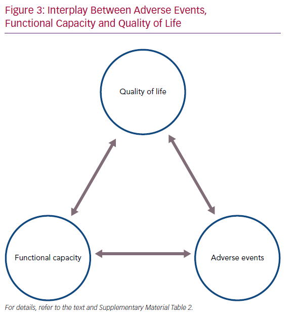
Interplay Between Adverse Events

Article image

Exercise capacity is closely linked to QoL. Better QoL is related to better muscular strength, treadmill time, anaerobic threshold and pVO2, all of which are factors describing aspects of FC.14,16,26 The fact that increasing FC is associated with better QoL in patients implanted with a continuous-flow LVAD, as in HF patients not supported by an LVAD, highlights the need for continued focus on optimising exercise tolerance.20 Indeed, an interplay between AEs, FC and QoL exists in LVAD recipients, and to improve overall QoL the other two components must be addressed (Figure 3).

Conclusion

Based on a literature review, it is clear that both FC and QoL are severely impaired in advanced HF patients prior to LVAD implantation, but significant improvements are observed after implantation, even though FC remains severely reduced after implantation. Important preoperative predictors of low FC are age, diabetes, COPD, INTERMACS profile, NYHA class, AF and baseline walking distance (e.g. the ability to perform an FC test at baseline). Importantly, poor FC after LVAD implantation is closely related to QoL and is associated with the risk of AEs. These factors should be considered when considering LVAD implantation, especially as destination therapy, and reversible modifiable factors should be aggressively managed both before and after LVAD implantation.

Click here to see Supplementary material.

References

  1. Goldstein DJ, Meyns B, Xie R, et al. Third Annual Report From the ISHLT Mechanically Assisted Circulatory Support Registry: a comparison of centrifugal and axial continuous-flow left ventricular assist devices. J Hear Lung Transplant. 2019;38:352-363.
    Crossref | PubMed
  2. de By TMMH, Mohacsi P, Gahl B, et al. The European Registry for Patients with Mechanical Circulatory Support (EUROMACS) of the European Association for Cardio-Thoracic Surgery (EACTS): second report. Eur J Cardiothorac Surg 2018;53:309–16.
    Crossref | PubMed
  3. Nakatani T, Sase K, Oshiyama H, et al. Japanese registry for Mechanically Assisted Circulatory Support: first report. J Heart Lung Transplant 2017;36:1087–96.
    Crossref | PubMed
  4. Braun O, Nilsson J, Gustafsson F, et al. Continuous-flow LVADs in the Nordic countries: complications and mortality and its predictors. Scand Cardiovasc J 2019;53:14–20.
    Crossref | PubMed
  5. Mehra MR, Uriel N, Naka Y, et al. A fully magnetically levitated left ventricular assist device – final report. N Engl J Med 2019;380:1618–27.
    Crossref | PubMed
  6. Mehra MR, Goldstein DJ, Uriel N, et al. Two-year outcomes with a magnetically levitated cardiac pump in heart failure. N Engl J Med 2018;378:1386–95.
    Crossref | PubMed
  7. Gustafsson F, Rogers JG. Left ventricular assist device therapy in advanced heart failure: patient selection and outcomes. Eur J Heart Fail 2017;19:595–602.
    Crossref | PubMed
  8. Saeed D, Garbade J, Gustafsson F, et al. Two-year outcomes in real world patients treated with Heartmate 3™ left ventricular assist device for advanced heart failure: data from the ELEVATE registry. J Heart Lung Transplant 2019;38(Suppl):S67.
    Crossref
  9. Rogers JG, Aaronson KD, Boyle AJ, et al. Continuous flow left ventricular assist device improves functional capacity and quality of life of advanced heart failure patients. Am J Coll Cardiol 2010;55:1826–34.
    Crossref | PubMed
  10. Kirklin JK, Pagani FD, Kormos RL, et al. Eighth annual INTERMACS report: special focus on framing the impact of adverse events. J Heart Lung Transplant 2017;36:1080–6.
    Crossref | PubMed
  11. Jung MH, Gustafsson F. Exercise in heart failure patients supported with a left ventricular assist device. J Hear Lung Transplant. 2015;34:489-496.
    Crossref | PubMed
  12. Mirza KK, Cuomo K, Jung MH, et al. Effect of heart rate reserve on exercise capacity in patients treated with a continuous flow left ventricular assist device. ASAIO J 2020;66:160–5.
    Crossref | PubMed
  13. Hayes K, Leet AS, Bradley SJ, Holland AE. Effects of exercise training on exercise capacity and quality of life in patients with a left ventricular assist device: a preliminary randomized controlled trial. J Heart Lung Transplant 2012;31:729–34.
    Crossref | PubMed
  14. Kerrigan DJ, Williams CT, Ehrman JK, et al. Muscular strength and cardiorespiratory fitness are associated with health status in patients with recently implanted continuous-flow LVADs. Cardiopulm Rehabil Prev 2013;33:396–400.
    Crossref | PubMed
  15. Jakovljevic DG, McDiarmid A, Hallsworth K, et al. Effect of left ventricular assist device implantation and heart transplantation on habitual physical activity and quality of life. Am J Cardiol 2014;114:88–93.
    Crossref | PubMed
  16. Kerrigan DJ, Williams CT, Ehrman JK, et al. Cardiac rehabilitation improves functional capacity and patient-reported health status in patients with continuous-flow left ventricular assist devices: the Rehab-VAD randomized controlled trial. JACC Heart Fail 2014;2:653–9.
    Crossref | PubMed
  17. Modica M, Ferratini M, Torri A, et al. Quality of life and emotional distress early after left ventricular assist device implant: a mixed-method study. Artif Organs 2015;39:220–7.
    Crossref | PubMed
  18. Kiernan MS, Sundareswaran KS, Pham DT, et al. Preoperative determinants of quality of life and functional capacity response to left ventricular assist device therapy. J Card Fail 2016;22:797–805.
    Crossref | PubMed
  19. Jung MH, Houston B, Russell SD, Gustafsson F. Pump speed modulations and sub-maximal exercise tolerance in left ventricular assist device recipients: a double-blind, randomized trial. J Heart Lung Transplant 2017;36:36–41.
    Crossref | PubMed
  20. Jung MH, Goetze JP, Boesgaard S, Gustafsson F. Neurohormonal activation and exercise tolerance in patients supported with a continuous-flow left ventricular assist device. Int J Cardiol 2016;220:196–200.
    Crossref | PubMed
  21. Dimopoulos S, Diakos N, Tseliou E, et al. Chronotropic incompetence and abnormal heart rate recovery early after left ventricular assist device implantation. Pacing Clin Electrophysiol 2011;34:1607–14.
    Crossref | PubMed
  22. Jakovljevic DG, Birks EJ, George RS, et al. Relationship between peak cardiac pumping capability and selected exercise-derived prognostic indicators in patients treated with left ventricular assist devices. Eur J Heart Fail 2011;13:992–9.
    Crossref | PubMed
  23. Jacquet L, Vancaenegem O, Pasquet A, et al. Exercise capacity in patients supported with rotary blood pumps is improved by a spontaneous increase of pump flow at constant pump speed and by a rise in native cardiac output. Artif Organs 2011;35:682–90.
    Crossref | PubMed
  24. Hasin T, Topilsky Y, Kremers WK, et al. Usefulness of the six-minute walk test after continuous axial flow left ventricular device implantation to predict survival. Am J Cardiol 2012;110:1322–8.
    Crossref | PubMed
  25. Noor MR, Bowles C, Banner NR. Relationship between pump speed and exercise capacity during HeartMate II left ventricular assist device support: Influence of residual left ventricular function. Eur J Heart Fail 2012;14:613–20.
    Crossref | PubMed
  26. Karapolat H, Engin C, Eroglu M, et al. Efficacy of the cardiac rehabilitation program in patients with end-stage heart failure, heart transplant patients, and left ventricular assist device recipients. Transplant Proc 2013;45:3381–5.
    Crossref | PubMed
  27. Grosman-Rimon L, McDonald MA, Pollock Bar-Ziv S, et al. Chronotropic incompetence, impaired exercise capacity, and inflammation in recipients of continuous-flow left ventricular assist devices. J Heart Lung Transplant 2013;32:930–2.
    Crossref | PubMed
  28. Martina J, Jonge N, Rutten M, et al. Exercise hemodynamics during extended continuous flow left ventricular assist device support: The response of systemic cardiovascular parameters and pump performance. Artif Organs 2013;37:754–62.
    Crossref | PubMed
  29. Camboni D, Lange TJ, Ganslmeier P, et al. Left ventricular support adjustment to aortic valve opening with analysis of exercise capacity. J Cardiothorac Surg 2014;9:1–7.
    Crossref | PubMed
  30. Imamura T, Kinugawa K, Nitta D, et al. Perioperative hypoalbuminemia affects improvement in exercise tolerance after left ventricular assist device implantation. Circ J 2015;79:1970–5.
    Crossref | PubMed
  31. Imamura T, Kinugawa K, Nitta D, et al. Opening of aortic valve during exercise is key to preventing development of aortic insufficiency during ventricular assist device treatment. ASAIO J 2015;61:514–19.
    Crossref | PubMed
  32. Marko C, Danzinger G, Käferbäck M, et al. Safety and efficacy of cardiac rehabilitation for patients with continuous flow left ventricular assist devices. Eur J Prev Cardiol 2015;22:1378–84.
    Crossref | PubMed
  33. Kerrigan DJ, Williams CT, Brawner CA, et al. Heart rate and VO2 concordance in continuous-flow left ventricular assist devices. Med Sci Sports Exerc 2016;48:363–7.
    Crossref | PubMed
  34. Jung MH, Houston B, Russell SD, Gustafsson F. Pump speed modulations and sub-maximal exercise tolerance in left ventricular assist device recipients: A double-blind, randomized trial. J Heart Lung Transplant 2017;36:36–41.
    Crossref | PubMed
  35. Fresiello L, Buys R, Timmermans P, et al. Exercise capacity in ventricular assist device patients: clinical relevance of pump speed and power. Eur J Cardiothorac Surg 2016;50:752–7.
    Crossref | PubMed
  36. Vignati C, Apostolo A, Cattadori G, et al. Lvad pump speed increase is associated with increased peak exercise cardiac output and vo2, postponed anaerobic threshold and improved ventilatory efficiency. Int J Cardiol 2017;230:28–32.
    Crossref | PubMed
  37. Marko C, Xhelili E, Lackner T, et al. Exercise performance during the first two years after left ventricular assist device implantation. ASAIO J 2017;63:408–13.
    Crossref | PubMed
  38. Lim HS, Howell N, Ranasinghe A. The effect of left ventricular assist device therapy in patients with heart failure and mixed pulmonary hypertension. Int J Artif Organs 2017;40:67–73.
    Crossref | PubMed
  39. Lairez O, Delmas C, Fournier P, et al. Feasibility and accuracy of gated blood pool SPECT equilibrium radionuclide ventriculography for the assessment of left and right ventricular volumes and function in patients with left ventricular assist devices. J Nucl Cardiol 2018;25:625–34.
    Crossref | PubMed
  40. Apostolo A, Paolillo S, Contini M, et al. Comprehensive effects of left ventricular assist device speed changes on alveolar gas exchange, sleep ventilatory pattern, and exercise performance. J Heart Lung Transplant 2018;37:1361–71.
    Crossref | PubMed
  41. Schmidt T, Bjarnason-Wehrens B, Bartsch P, et al. Exercise capacity and functional performance in heart failure patients supported by a left ventricular assist device at discharge from inpatient rehabilitation. Artif Organs 2018;42:22–30.
    Crossref | PubMed
  42. Mezzani A, Pistono M, Agostoni P, et al. Exercise gas exchange in continuous-flow left ventricular assist device recipients. PLoS One 2018;13:e0187112.
    Crossref | PubMed
  43. Rosenbaum AN, Dunlay SM, Pereira NL, et al. Determinants of improvement in cardiopulmonary exercise testing after left ventricular assist device implantation. ASAIO J 2018;64:610–5.
    Crossref | PubMed
  44. Racca V, Castiglioni P, Panzarino C, et al. Differences in biochemical markers between heart-transplanted and left ventricular assist device implanted patients, during cardiac rehabilitation. Sci Rep 2018;8:10816.
    Crossref | PubMed
  45. Schmidt T, Bjarnason-Wehrens B, Mommertz S, et al. Development of exercise-related values in heart failure patients supported with a left ventricular assist device. Int J Artif Organs 2019;42:201–6.
    Crossref | PubMed
  46. Gross C, Marko C, Mikl J, et al. LVAD pump flow does not adequately increase with exercise. Artif Organs 2019;43:222–8.
    Crossref | PubMed
  47. Koshy A, Cruz NB, Okwose NC, et al. Left ventricular filling pressures contribute to exercise limitation in patients with continuous flow left ventricular assist devices. ASAIO J 2020;247–52.
    Crossref | PubMed
  48. Gustafsson F, Mirza K, Pya Y, et al. Predictors of physical capacity six months after implantation of a full magnetically levitated left ventricular assist device: an analysis from the ELEVATE registry. J Card Fail 2020;26:580–7.
    Crossref | PubMed
  49. Cowger JA, Naka Y, Aaronson KD, et al. Quality of life and functional capacity outcomes in the MOMENTUM 3 trial at 6 months: a call for new metrics for left ventricular assist device patients. J Heart Lung Transplant 2018;37:15–24.
    Crossref | PubMed
  50. Jung MH, Hansen PB, Sander K, et al. Effect of increasing pump speed during exercise on peak oxygen uptake in heart failure patients supported with a continuous-flow left ventricular assist device. A double-blind randomized study. Eur J Heart Fail 2014;16:403–8.
    Crossref | PubMed
  51. Mehra MR, Naka Y, Uriel N, et al. A fully magnetically levitated circulatory pump for advanced heart failure. N Engl J Med 2017;376:440–50.
    Crossref | PubMed
  52. Netuka I, Sood P, Pya Y, et al. Fully magnetically levitated left ventricular assist system for treating advanced HF: a multicenter study. J Am Coll Cardiol 2015;66:2579–89.
    Crossref | PubMed
  53. Miller LW, Pagani FD, Russell SD, et al. Use of a continuous-flow device in patients awaiting heart transplantation. N Engl J Med 2007;357:885–96.
    Crossref | PubMed
  54. Krabatsch T, Netuka I, Schmitto JD, et al. Heartmate 3 fully magnetically levitated left ventricular assist device for the treatment of advanced heart failure – 1 year results from the Ce mark trial. J Cardiothorac Surg 2017;12:23.
    Crossref | PubMed
  55. Slaughter MS, Rogers JG, Milano CA, et al. Advanced heart failure treated with continuous-flow left ventricular assist device. N Engl J Med 2009;361:2241–51.
    Crossref | PubMed
  56. Aaronson KD, Slaughter MS, Miller LW, et al. Use of an intrapericardial, continuous-flow, centrifugal pump in patients awaiting heart transplantation. Circulation 2012;125:3191–200.
    Crossref | PubMed
  57. Starling RC, Estep JD, Horstmanshof DA, et al. Risk assessment and comparative effectiveness of left ventricular assist device and medical management in ambulatory heart failure patients: the ROADMAP study 2-year results. JACC Heart Fail 2017;5:518–27.
    Crossref | PubMed
  58. Gustafsson F, Shaw S, Lavee J, et al. Six-month outcomes after treatment of advanced heart failure with a full magnetically levitated continuous flow left ventricular assist device: report from the ELEVATE registry. Eur Heart J 2018;39:3454–60.
    Crossref | PubMed
  59. Rogers JG, Pagani FD, Tatooles AJ, et al. Intrapericardial left ventricular assist device for advanced heart failure. N Engl J Med 2017;376:451–60.
    Crossref | PubMed
  60. Milano CA, Rogers JG, Tatooles AJ, et al. HVAD: the ENDURANCE supplemental trial. JACC Heart Fail 2018;6:792–802.
    Crossref | PubMed
  61. Jung MH, Gustafsson F, Houston B, Russell SD. Ramp study hemodynamics, functional capacity, and outcome in heart failure patients with continuous-flow left ventricular assist devices. ASAIO J 2016;62:442–6.
    Crossref | PubMed
  62. Imamura T, Kinugawa K, Nitta D, et al. Opening of native aortic valve accomplished after left ventricular assist device implantation in patients with insufficient preoperative beta-blocker treatment. Int Heart J 2015;56:303–8.
    Crossref | PubMed
  63. Mirza KK, Jung MH, Sigvardsen PE, et al. Computed tomography – estimated right ventricular function and exercise capacity in patients with continuous-flow left ventricular assist devices. ASAIO J 2020;66:8–16.
    Crossref | PubMed
  64. Cohn JN, Johnson G, Ziesche S, et al. A comparison of enalapril with hydralazine-isosorbide dinitrate in the treatment of chronic congestive heart failure. N Engl J Med 1991;325:303–10.
    Crossref | PubMed
  65. Young JB, Abraham WT, Smith AL, et al. Combined cardiac resynchronization and implantable cardioversion defibrillation in advanced chronic heart failure. JAMA 2003;289:2685.
    Crossref | PubMed
  66. Arnold S V, Jones PG, Allen LA, et al. Frequency of poor outcome (death or poor quality of life) after left ventricular assist device for destination therapy. Circ Heart Fail 2016;9:e002800.
    Crossref | PubMed
  67. Murninkas D, Alba AC, Delgado D, et al. Right ventricular function and prognosis in stable heart failure patients. J Card Fail 2014;20:343–9.
    Crossref | PubMed
  68. Villela MA, Guerrero-Miranda CY, Chinnadurai T, et al. Rate response pacing in left ventricular assist device patients. ASAIO J 2020:E29–30.
    Crossref | PubMed
  69. Muthiah K, Robson D, Prichard R, et al. Effect of exercise and pump speed modulation on invasive hemodynamics in patients with centrifugal continuous-flow left ventricular assist devices. J Heart Lung Transplant 2015;34:522–9.
    Crossref | PubMed
  70. Butler J, Howser R, Portner PM, Pierson RN. Body mass index and outcomes after left ventricular assist device placement. Ann Thorac Surg 2005;79:66–73.
    Crossref | PubMed
  71. Go PH, Nemeh HW, Borgi J, et al. Effect of body mass index on outcomes in left ventricular assist device recipients. J Card Surg 2016;31:242–7.
    Crossref | PubMed
  72. Rashid MA, Qureshi BA, Ahmed N, Sherwani MA. Impact of body mass index on left ventricular mass. J Ayub Med Coll Abbottabad 2014;26:167–9.
    PubMed
  73. Clerkin KJ, Naka Y, Mancini DM, et al. The impact of obesity on patients bridged to transplantation with continuous-flow left ventricular assist devices. JACC Heart Fail 2016;4:761–8.
    Crossref | PubMed
  74. Amione-Guerra J, Cruz-Solbes AS, Bhimaraj A, et al. Anemia after continuous-flow left ventricular assist device implantation: characteristics and implications. Int J Artif Organs 2017;40:481–8.
    Crossref | PubMed
  75. Bode LE, Wesner S, Katz JN, et al. Intravenous versus oral iron replacement in patients with a continuous-flow left ventricular assist device. ASAIO J 2019;65:E90–1.
    Crossref | PubMed
  76. Amione-Guerra J, Cruz-Solbes S, Bhimaraj A, et al. Iron deficiency is the most common cause of anemia in CF-LVAD patients. Int J Artif Organs 2017;40:481–8.
    Crossref | PubMed
  77. Mardis A, Robinson C, Stafford B, et al. Intravenous iron replacement in patients with left ventricular assist devices. A pilot study. J Heart Lung Transplant 2019;38(Suppl):S112–13.
    PubMed
  78. Lim HS, Howell N, Ranasinghe A. The physiology of continuous-flow left ventricular assist devices. J Card Fail 2017;23:169–80.
    Crossref | PubMed
  79. Grosman-Rimon L, Kachel E, McDonald MA, et al. Association between neurohormone levels and exercise testing measures in patients with mechanical circulatory supports. ASAIO J 2019.
    Crossref | PubMed
  80. Camboni D, Lange TJ, Ganslmeier P, et al. Left ventricular support adjustment to aortic valve opening with analysis of exercise capacity. J Cardiothorac Surg 2014;9:93.
    Crossref | PubMed
  81. Imamura T, Kinugawa K, Nitta D, et al. Novel scoring system using postoperative cardiopulmonary exercise testing predicts future explantation of left ventricular assist device. Circ J 2015;79:560–6.
    Crossref | PubMed
  82. Schmidt T, Bjarnason-Wehrens B, Mommertz S, et al. Changes in total cardiac output and oxygen extraction during exercise in patients supported with an HVAD left ventricular assist device. Artif Organs 2018;42:22–30.
    Crossref | PubMed
  83. Majani G, Giardini A, Opasich C, et al. Effect of valsartan on quality of life when added to usual therapy for heart failure: results from the Valsartan Heart Failure trial. J Card Fail 2005;11:253–9.
    Crossref | PubMed
  84. Spertus J, Peterson E, Conard MW, et al. Monitoring clinical changes in patients with heart failure: A comparison of methods. Am Heart J 2005;150:707–15.
    Crossref | PubMed
  85. Rector TS, Cohn JN. Assessment of patient outcome with the Minnesota Living with Heart Failure questionnaire: Reliability and validity during a randomized, double-blind, placebo-controlled trial of pimobendan. Am Heart J 1992;124:1017–25.
    PubMed